
Q. Is lamina dura radiopaque?
The lamina dura surrounds the tooth socket and provides the attachment surface with which the Sharpey’s fibers of the periodontal ligament perforate. On an x-ray a lamina dura will appear as a radiopaque line surrounding the tooth root. An intact lamina dura is seen as a sign of healthy periodontium.
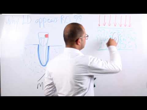
Q. What is lamina dura in radiograph?
Lamina dura (LD) is a radiographic landmark viewed largely on periapical radiographs (PR). The terminology LD (or alveolus) is applied to the thin layer of dense cortical bone, which lines the roots of sound teeth. Presence of LD is an indication of the health of the teeth.
Table of Contents
- Q. Is lamina dura radiopaque?
- Q. What is lamina dura in radiograph?
- Q. Is lamina dura the same as bundle bone?
- Q. What does loss of lamina dura indicate?
- Q. Is lamina dura cribriform plate?
- Q. Which term is also used to refer to the lamina dura?
- Q. What is lamina dura?
- Q. What is fenestration and dehiscence?
- Q. What is PDL widening?
- Q. How can you tell Denestration and dehiscence?
- Q. What is tooth fenestration?
- Q. What is the lamina dura?
- Q. What do you need to know about lamina dura?
- Q. Is the lamina dura an irregular radiolucent line?
- Q. Is the loss of the apical lamina dura a radiographic sign?
- Q. Why are the walls of the lamina dura called cribriform plate?
Q. Is lamina dura the same as bundle bone?
The alveolar process includes a region of compact bone that is adjacent to the periodontal ligament (PDL). This is called the lamina dura when it is viewed on radiographs. The alveolar bone proper is also called the bundle bone because of the Sharpey fibers.
Q. What does loss of lamina dura indicate?
It is usually considered that the loss of the lamina dura is pathognomonic of hyperparathyroidism although some degree of loss may be apparent in osteomalacia and in Paget’s disease.
Q. Is lamina dura cribriform plate?
The lamina dura is produced by the periodontal ligament; it forms the hard lining of the tooth socket, and is a cribriform plate of bundle bone in which fibers of the periodontal ligament are em- bedded.
Q. Which term is also used to refer to the lamina dura?
: the thin hard layer of bone that lines the socket of a tooth and that appears as a dense white line in radiography. — called also cribriform plate.
Q. What is lamina dura?
Medical Definition of lamina dura : the thin hard layer of bone that lines the socket of a tooth and that appears as a dense white line in radiography. — called also cribriform plate.
Q. What is fenestration and dehiscence?
Fenestration is the condition, in which the bony coverage of the root surface is lost, and the root surface is only covered by the periosteum and gingiva. In such lesions, marginal bone is intact. When this bone defect spreads toward the marginal bone, it is called dehiscence.[1]
Q. What is PDL widening?
PDL widening, thickening of the lamina dura, increased number and size of trabeculae and bone loss are radiographic features. Continuous force causes tooth movement that is marked initially by PDL narrowing. In the secondary period of tooth movement, the PDL is considerably widened.
Q. How can you tell Denestration and dehiscence?
Fenestration was considered as a local bone defect or as bone exposure of overlying alveolar bone on the root surface with the intact marginal bone. When the bone defect spread to the marginal bone, this was considered to be dehiscence.
Q. What is tooth fenestration?
Fenestration is an isolated area in which the tooth root is denuded of bone and the root surface is covered only by periosteum and overlying gingiva. Mucosal fenestration is a clinical entity in which the overlying gingiva or mucosa is also denuded thus the root is exposed to the oral cavity.
Q. What is the lamina dura?
The lamina dura is the hard bony lining of the alveolus (Fig. 1), or socket, of a tooth, and to- gether with the periodontal ligament and the cementum which coats the tooth root it forms the attachment apparatus of the tooth.
Q. What do you need to know about lamina dura?
Introduction Lamina dura (LD) is a radiographic landmark viewed largely on periapical radiographs (PR). The terminology LD (or alveolus) is applied to the thin layer of dense cortical bone, which lines the roots of sound teeth. Presence of LD is an indication of the health of the teeth.
Q. Is the lamina dura an irregular radiolucent line?
The lamina dura is a/an regular radiolucent line around the roots. irregular radiolucent line around the roots.
Q. Is the loss of the apical lamina dura a radiographic sign?
Another early radiographic sign is loss of the apical lamina dura (Figure 6-4, B–D). This is also not a reliable indicator of disease because the apical lamina dura frequently becomes unidentifiable when there is insufficient overlying radiodense tissue.
Q. Why are the walls of the lamina dura called cribriform plate?
They should also be well positioned to avoid elongation, foreshortening, angulation, or distortion of the image. The compact bone that forms the walls of the alveolus (the white line of the lamina dura on radiographs) is also referred to as a cribriform plate due to its multiple perforations for vessels and nerves.