
Horizontal cells form feedback synapses with cones and feedforward synapses with CBCs. However, the exact computational role of HCs is still debated. Along with performing global signaling within their laterally coupled network, HCs also provide local, cone-specific feedback.
Q. How do horizontal cells work?
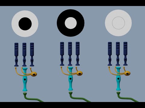
Horizontal cells receive input from multiple photoreceptor cells. They use that input to integrate signaling from different populations of photoreceptor cells, make adjustments to the signals that will be sent to bipolar cells, and regulate activity in photoreceptor cells themselves.
Table of Contents
- Q. How do horizontal cells work?
- Q. What do horizontal and amacrine cells do?
- Q. What is the function of ganglion cells?
- Q. Do horizontal cells release GABA?
- Q. Are horizontal cells GABAergic?
- Q. Do horizontal cells fire action potentials?
- Q. What is a bipolar cell?
- Q. Where are bipolar cells found?
- Q. What is the difference between ON and OFF bipolar cells?
- Q. Do bipolar cells generate action potentials?
- Q. Which neurons are bipolar?
- Q. Do amacrine cells fire action potentials?
- Q. How is the rod off channel generated?
- Q. Can Rod cells detect color?
- Q. Do photoreceptors depolarize in the presence of light?
- Q. Does light depolarize or Hyperpolarize photoreceptors?
- Q. What are bipolar and ganglion cells?
- Q. What happens when a rod is stimulated by light?
- Q. How are rods activated?
- Q. Which structure controls the amount of light entering the eye?
- Q. What happens when a photon strikes rhodopsin?
- Q. How light is converted to an action potential?
- Q. What is the ultimate higher order olfactory destination?
- Q. What is Metarhodopsin II?
Q. What do horizontal and amacrine cells do?
The horizontal cells receive information from the photoreceptors and transmit it to a number of surrounding bipolar neurons. The amacrine cells receive their inputs from the bipolar cells and do the same thing to the ganglion neurons: activate the ones that are in their vicinity.
Q. What is the function of ganglion cells?
Introduction. Ganglion cells are the projection neurons of the vertebrate retina, conveying information from other retinal neurons to the rest of the brain. Their perikarya are the largest of any retinal neurons and are located along the inner margin of the retina, in the ganglion cell layer.
Q. Do horizontal cells release GABA?
Functional properties Horizontal cells are depolarized by the release of glutamate from photoreceptors, which happens in the absence of light. Depolarization of a horizontal cell causes it to release the inhibitory neurotransmitter GABA on an adjacent photoreceptor.
Q. Are horizontal cells GABAergic?
Mouse horizontal cells are thus atypical GABAergic neurons, with no functional GABA uptake, but a glutamate and/or glutamine transport system allowing GABA synthesis, probably depending physiologically from glutamate released by photoreceptors.
Q. Do horizontal cells fire action potentials?
The same is true for retinal bipolar cells, horizontal cells, and many amacrine cells, which are equally tiny. The cells of the outer retina do not need to make action potentials, because electrotonic decrements are very small. In contrast, the retinal ganglion cell must send an axon several centimeters to the brain.
Q. What is a bipolar cell?
Bipolar cells are interneurons in the retina ( Vision), which transfer visual information from photoreceptors (rods and cones; Photoreceptors) to amacrine ( Retinal direction selectivity: Role of starburst amacrine cells) and ganglion cells ( Retinal ganglion cells).
Q. Where are bipolar cells found?
Often found in the retina, bipolar cells are crucial as they serve as both direct and indirect cell pathways. The specific location of the bipolar cells allow them to facilitate the passage of signals from where they start in the receptors to where they arrive at the amacrine and ganglion cells.
Q. What is the difference between ON and OFF bipolar cells?
ON-center bipolar cells are depolarized by small spot stimuli positioned in the receptive field center. OFF-center bipolar cells are hyperpolarized by the same stimuli. Both types are repolarized by light stimulation of the peripheral receptive field outside the center (Fig. 1).
Q. Do bipolar cells generate action potentials?
Scientists have now been able to show that already bipolar cells can generate “digital” signals. At least three types of mouse BC showed clear evidence of fast and stereotypic action potentials, so called “spikes”.
Q. Which neurons are bipolar?
Bipolar cells (BCs) are the central neurons of the retina which carry light-elicited signals from photoreceptors and horizontal cells (HCs) in the outer retina to amacrine cells (ACs) and ganglion cells (GCs) in the inner retina.
Q. Do amacrine cells fire action potentials?
Amacrine cells are the first neurons in the visual system to fire action potentials, and also the first to generate transient responses. They send processes laterally along the inner plexiform layer, at the level of the bipolar-to-ganglion cell synapse (Figure 1).
Q. How is the rod off channel generated?
The ON- and OFF-channels in the mammalian retina are generated by cone photoreceptors connecting to several subtypes of ON- and OFF-cone bipolar cells and by rod photoreceptors connecting to one type of ON-rod bipolar cell. The ON- and OFF-type bipolar cells express functionally different types of glutamate receptors.
Q. Can Rod cells detect color?
These specialized cells are called photoreceptors. There are 2 types of photoreceptors in the retina: rods and cones. The rods are most sensitive to light and dark changes, shape and movement and contain only one type of light-sensitive pigment. Rods are not good for color vision.
Q. Do photoreceptors depolarize in the presence of light?
In humans and other vertebrates, neurotransmitter release occurs in the dark (when the photoreceptor plasma membrane is depolarized). In the presence of light, however, the cell becomes hyperpolarized, and neurotransmitter release is inhibited.
Q. Does light depolarize or Hyperpolarize photoreceptors?
The retina has many layers of various cell types. The most numerous photoreceptor cells (rods and cones) form the outermost layer. Unlike most sensory receptor cells, photoreceptors actually become hyperpolarized when stimulated; and conversely are depolarized when not stimulated.
Q. What are bipolar and ganglion cells?
As a part of the retina, bipolar cells exist between photoreceptors (rod cells and cone cells) and ganglion cells. They act, directly or indirectly, to transmit signals from the photoreceptors to the ganglion cells.
Q. What happens when a rod is stimulated by light?
The retinal exists in the 11-cis-retinal form when in the dark, and stimulation by light causes its structure to change to all-trans-retinal. This structural change causes an increased affinity for the regulatory protein called transducin (a type of G protein).
Q. How are rods activated?
Publisher Summary. In vertebrate retinal rod cells, the absorption of a photon of light by rhodopsin triggers the activation of a G protein. Several hundred G proteins are activated by one molecule of photoexcited receptor. To terminate the signal, rhodopsin must be inactivated.
Q. Which structure controls the amount of light entering the eye?
Iris
Q. What happens when a photon strikes rhodopsin?
What happens when a photon strikes rhodopsin? – Neurotransmitter release increases. – Entry of sodium ions into cells accelerates. – Rhodopsin is degraded and is not useful.
Q. How light is converted to an action potential?
The structure of the eye responsible for converting light waves into action potentials is the retina. The neural layer of the retina is composed of three main types of cells: the photoreceptors, the bipolar neurons and the ganglion cells.
Q. What is the ultimate higher order olfactory destination?
What is the ultimate higher-order olfactory destination? the olfactory cortex. What skull structure must the axons from the olfactory epithelium pass through in order to get to the olfactory bulbs? cribriform plate.
Q. What is Metarhodopsin II?
The product of light activation, Metarhodopsin II, initiates the visual phototransduction pathway by stimulating the G protein transducin (Gt), resulting in the liberation of its α subunit. This GTP-bound subunit in turn activates cGMP phosphodiesterase.